Vaginitis in Focus | Part 2: Aerobic Vaginitis Beyond Pathogen Detection
Improving diagnosis and clinical management through evidence of inflammation, epithelial change and altered vaginal flora.
A positive bacterial result can appear to offer a straightforward explanation for persistent vaginal symptoms. For aerobic vaginitis (AV), however, identifying an organism is only part of the clinical question. The decision to treat also depends on whether the examination and microscopic findings support an inflammatory vaginal disorder.
The first article in this series examined inappropriate anti-infective use. Here, AV illustrates a practical challenge behind that problem: how to distinguish a clinically meaningful syndrome from a laboratory finding, and how to turn that distinction into more appropriate management.
Why AV needs its own diagnostic reasoning
The original description of AV drew on an observational study of 631 women attending prenatal care or a vaginitis clinic in Leuven, Belgium. It distinguished an abnormal vaginal flora pattern involving reduced lactobacilli, altered bacterial morphotypes and inflammatory or epithelial changes from bacterial vaginosis (Donders et al., 2002).
This distinction has practical consequences. Donders et al. (2015) emphasize that AV can be confused with BV or intermediate Nugent findings, despite differences in the inflammatory and epithelial components. A BV-oriented result cannot, on its own, resolve every question about suspected AV. Likewise, a culture growing an enteric organism does not establish that the organism explains the patient's symptoms.
The European guideline explicitly separates culture positivity from AV diagnosis. Culture and susceptibility testing may assist treatment in selected circumstances, but culture is not recommended as the diagnostic basis for AV (Sherrard et al., 2023).
The clinical priority is to establish a compatible disorder before selecting an antimicrobial, rather than allowing a positive organism result to define the disorder.
Make the microscopic assessment reproducible
Wet-mount examination provides information about host cells as well as bacterial morphotypes. The ISSVD microscopy guideline addresses sampling, preparation, systematic reading and classification, emphasizing that interpretation requires training rather than simply access to a microscope (Vieira-Baptista et al., 2021).
For AV, a structured assessment considers lactobacillary grade, background flora, leukocyte abundance, the proportion of toxic leukocytes and parabasal epithelial cells. The ISSVD recommendations identify wet-mount microscopy, preferably with phase contrast, as the diagnostic reference and allow the AV score to describe severity (Vieira-Baptista et al., 2023).
For laboratories, the operational implication is to define what must be assessed and how uncertainty is communicated. A report that records an organism without the relevant cellular context may leave the clinician with an incomplete explanation. A report that identifies the method, findings and interpretive limits provides a more useful basis for discussion.
A prospective study of an alternative diagnostic approach

Title excerpt from Dong et al. (2022), p. 1. Read the paper.
Dong et al. (2022) developed criteria combining Gram-stained microscopy with clinical features at Tianjin Medical University General Hospital. The development group comprised 325 AV cases and 325 controls; 500 consecutive outpatients formed a separate prospective validation group. This was a single-center study with international coauthors, not a multinational trial.

Figure 1. Prospective validation against wet-mount reference classification, including single and mixed AV. Sensitivity and specificity use different denominators. Original visualization of Table 4 in Dong et al. (2022).
In the full validation analysis, sensitivity was 82.30% (93/113) and specificity 94.83% (367/387). The remaining 20 reference-positive cases were missed and 20 reference-negative cases classified positive. These results concern diagnostic agreement, not improved treatment outcomes (Dong et al., 2022).
The distinction between identifying cases and improving care matters. A promising diagnostic method still needs an implementation pathway: who reviews discordant findings, how results reach the clinician and what happens when symptoms and the laboratory interpretation do not align?
Coexisting findings complicate an AV diagnosis
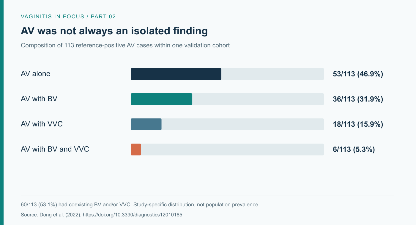
Figure 2. Composition of the 113 reference-positive AV cases within the validation cohort. Counts are reported by Dong et al. (2022), section 3.4.1; percentages are calculated. This is a study-specific case distribution, not a population prevalence estimate.
The reference-positive group included 53 single AV cases and 60 cases with BV and/or VVC. This illustrates why identifying one condition should not automatically close the diagnostic assessment (Dong et al., 2022).
For the clinician, coexisting findings create a prioritization problem: which abnormalities best explain the symptoms, which require treatment and which need further clarification? Simply adding medicines for every positive result risks replacing diagnostic reasoning with a longer prescription.
The European guideline describes mixed infections as frequent and recognizes persistent or recurrent symptoms as reasons for follow-up. These recommendations support reassessment when the initial explanation does not account for the patient's course (Sherrard et al., 2023).
Recognize the limits of the method
The proposed Gram-stain criteria do not reproduce every wet-mount feature: toxic leukocytes could not be assessed reliably, and clinical features were incorporated instead. The two scoring systems have different thresholds and should not be interchanged. Independent external validation and patient-outcome studies remain important (Dong et al., 2022).
Importantly, the 2023 ISSVD recommendations continue to use wet-mount microscopy as the reference and do not endorse routine Gram staining as a validated replacement for AV/DIV diagnosis (Vieira-Baptista et al., 2023). A locally evaluated alternative should therefore be described by its actual method and evidence, without implying universal guideline adoption.
Connect the diagnosis to a management plan
Donders et al. (2015) describe a treatment rationale that addresses the bacterial, inflammatory and atrophic components according to the findings. This helps explain why repeated antimicrobial courses alone may fail to address the dominant clinical problem. It does not mean that every patient should receive all treatment components.
The European guideline lists topical clindamycin for uncomplicated AV and additional approaches where inflammation or atrophy is prominent; its uncomplicated-AV recommendation is based on grade C evidence. Treatment selection must account for the individual presentation, contraindications and local guidance (Sherrard et al., 2023).
The ISSVD recommendations likewise acknowledge that much AV/DIV management advice rests on limited observational evidence and expert opinion. They do not identify metronidazole as a preferred AV/DIV treatment (Vieira-Baptista et al., 2023). Persistent symptoms after a presumed BV regimen should prompt reconsideration of the diagnosis, rather than an automatic extension of the same treatment.
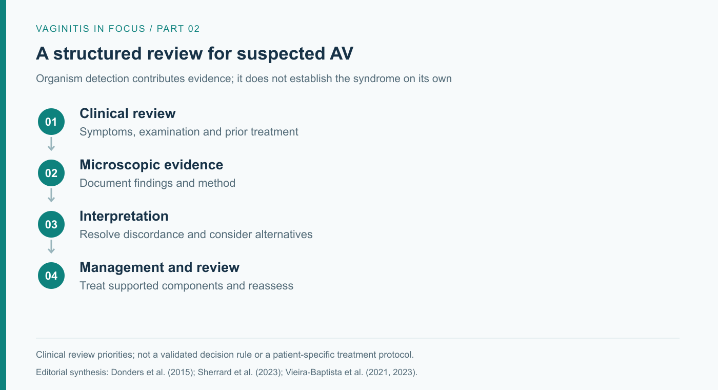
Figure 3. Editorial synthesis of clinical review priorities, informed by Donders et al. (2015), Sherrard et al. (2023) and Vieira-Baptista et al. (2021, 2023). This is not a prospectively validated clinical algorithm.
In practice, clinical and laboratory teams can organize the review around four questions:
- Does the presentation support AV? Revisit symptoms, examination findings, hormonal context and prior treatment, alongside the microscopic assessment.
- Could another diagnosis explain the findings? Severe inflammatory presentations warrant appropriate exclusion of trichomoniasis; noninfectious and atrophic disorders may also require consideration (Vieira-Baptista et al., 2023).
- Which component needs treatment? Document the rationale for antimicrobial, anti-inflammatory or atrophy-directed management, rather than treating an organism name in isolation.
- What will trigger reassessment? Agree on review when symptoms persist, worsen or recur. Reconsider the diagnosis and the quality of the evidence before repeating a prescription.
AV and desquamative inflammatory vaginitis (DIV) overlap in professional recommendations, particularly in severe presentations. They should not be treated as interchangeable labels for every patient, and outcomes from a DIV cohort should not be presented as outcomes for all AV.
A clinically useful diagnosis changes the next decision
Better AV management begins with a more precise question: do the clinical and microscopic findings explain this patient's symptoms, and what treatment do those findings justify? That question makes organism detection useful without allowing it to become the whole diagnosis.
For clinicians and laboratories, the immediate opportunity is to make the assessment reproducible, communicate uncertainty and review the patient's response. Precision lies in matching the management decision to the evidence available for that patient.
References
Donders, G. G. G., Ruban, K., & Bellen, G. (2015). Selecting anti-microbial treatment of aerobic vaginitis. Current Infectious Disease Reports, 17, Article 24. https://doi.org/10.1007/s11908-015-0477-6
Donders, G. G. G., Vereecken, A., Bosmans, E., Dekeersmaecker, A., Salembier, G., & Spitz, B. (2002). Definition of a type of abnormal vaginal flora that is distinct from bacterial vaginosis: Aerobic vaginitis. BJOG: An International Journal of Obstetrics & Gynaecology, 109(1), 34–43. https://doi.org/10.1111/j.1471-0528.2002.00432.x
Dong, M., Wang, C., Li, H., Yan, Y., Ma, X., Li, H., Li, X., Wang, H., Zhang, Y., Qi, W., Meng, K., Tian, W., Wang, Y., Fan, A., Han, C., Donders, G. G. G., & Xue, F. (2022). Aerobic vaginitis diagnosis criteria combining Gram stain with clinical features: An establishment and prospective validation study. Diagnostics, 12(1), Article 185. https://doi.org/10.3390/diagnostics12010185
Sherrard, J., Wilson, J., Donders, G., Mendling, W., & Jensen, J. S. (2023). 2018 European (IUSTI/WHO) guideline on the management of vaginal discharge (2023 revision). International Union Against Sexually Transmitted Infections. https://www.bashh.org/_userfiles/pages/files/iustivaginaldischargeguidelines_2023.pdf
Vieira-Baptista, P., Grincevičienė, Š., Oliveira, C., Fonseca-Moutinho, J., Cherey, F., & Stockdale, C. K. (2021). The International Society for the Study of Vulvovaginal Disease vaginal wet mount microscopy guidelines: How to perform, applications, and interpretation. Journal of Lower Genital Tract Disease, 25(2), 172–180. https://doi.org/10.1097/LGT.0000000000000595
Vieira-Baptista, P., Stockdale, C. K., & Sobel, J. (Eds.). (2023). International Society for the Study of Vulvovaginal Disease recommendations for the diagnosis and treatment of vaginitis. Ad Médic. https://doi.org/10.59153/adm.rdtv.001
